
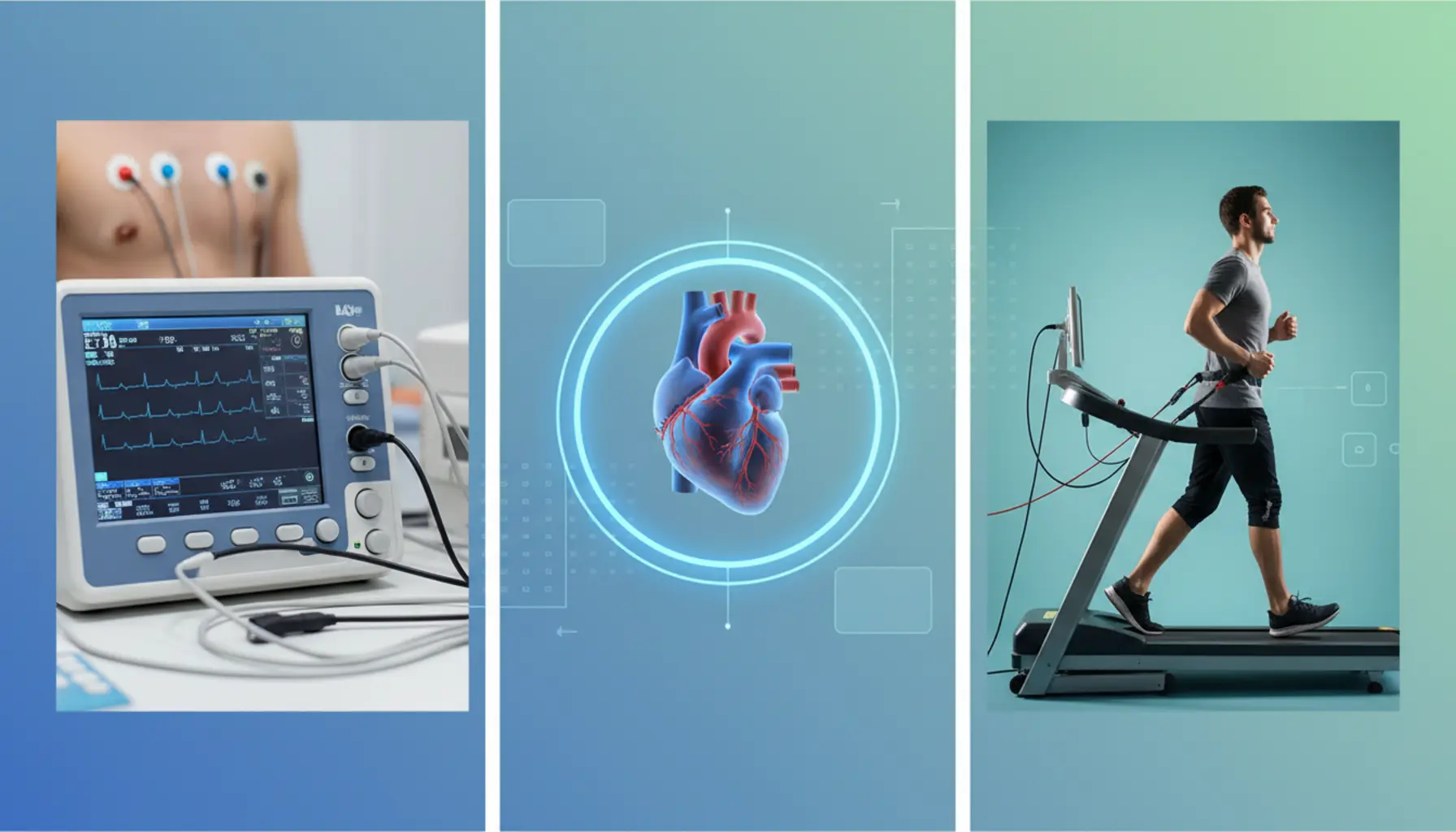
The exercise stress test ECG is a specialized diagnostic method that helps to detect heart diseases before the first symptoms appear. Thanks to this test, it is possible to comprehensively assess the function of the heart muscle under conditions of controlled exertion. Regular exercise ECGs allow you to take care of your heart’s health and safety.
Table of Contents
- What Is an Exercise Stress Test ECG?
- Why Is the Test Performed?
- Preparation for the Stress Test
- Step-by-Step: How the Test Is Conducted
- When Should the Stress Test Be Interrupted?
- Interpretation of ECG Results
What Is an Exercise Stress Test ECG?
The exercise stress test ECG, also known as a treadmill test or an exercise ECG, is a specialized cardiological examination that allows for the assessment of the heart’s function under conditions of controlled physical exertion. Unlike a standard resting ECG, which records the electrical activity of the heart in a non-moving state, the exercise test shows how the heart muscle performs when its oxygen demands increase—in other words, when your heart rate and breathing speed up and your body is “forced” into greater exertion. The idea behind the examination is simple: most problems related to ischemic heart disease or arrhythmias manifest themselves during effort rather than at rest, so the doctor deliberately simulates a load similar to brisk walking, climbing stairs, or intense walking. During the test, not only is the electrical activity of the heart monitored (recorded as an ECG tracing), but also heart rate, blood pressure, the patient’s well-being, and any possible symptoms such as chest pain, shortness of breath, dizziness, palpitations, or excessive fatigue. This enables the physician to obtain a much fuller and more dynamic picture of how the circulatory system functions compared to classic examinations performed at rest.
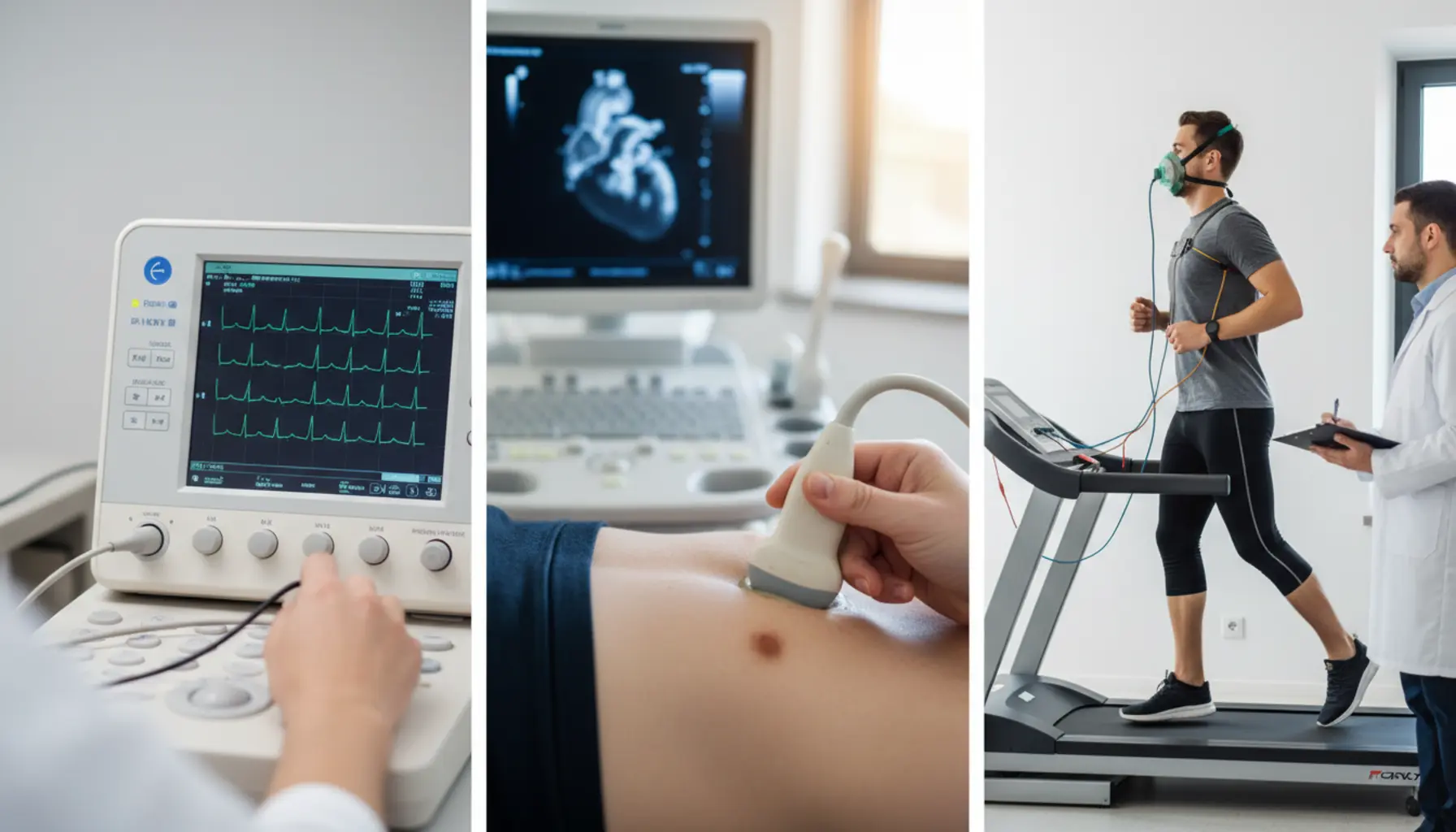
From a technical perspective, the exercise stress test ECG is most often performed by walking or running on a treadmill or pedaling a stationary bicycle (cycle ergometer), with a gradually increasing level of load. Before the test begins, a technician or nurse attaches several electrodes to the patient’s chest, connected to the ECG device to record cardiac activity in real time; a blood pressure cuff is placed on the arm, and the entire test is conducted in the presence of a doctor, ready to immediately interrupt the exercise if any alarming symptoms or changes are observed in the ECG. The load is usually increased every few minutes according to a set protocol (such as the Bruce protocol), which means that each phase brings a faster speed and steeper incline for the treadmill or greater resistance on the ergometer, with the goal being to reach so-called submaximal or maximal heart rate appropriate to the patient’s age and health status. In practice, the test is ended when the planned heart rate is reached, serious changes appear in the ECG tracing, blood pressure increases abnormally, excessive fatigue sets in, or the patient reports symptoms that cannot be ignored. The entire test record is then thoroughly analyzed—physicians mainly look for signs of myocardial ischemia (characteristic ST-segment depressions or elevations, changes in T waves), rhythm disturbances (e.g., tachycardia, extrasystoles), abnormal blood pressure and heart rate responses to exercise, and determine at what load level any abnormalities occur. Therefore, this examination helps detect coronary artery disease, assess the severity of already diagnosed heart conditions, monitor the efficacy of pharmacological or post-procedural treatment (such as after angioplasty or coronary artery bypass grafting), and determine what physical activity level is safe for a particular individual. The exercise ECG is a non-invasive, relatively short test (usually 15–30 minutes), performed under constant medical supervision, making it both a practical and safe diagnostic tool in everyday cardiological practice.
Why Is the Test Performed?
The exercise stress test ECG is mainly performed to assess how the heart copes with increased oxygen demand, i.e., in situations most similar to everyday physical activity. At rest, the heart muscle can be “silent” and not reveal signs of ischemia or arrhythmias, which only become apparent during exertion. The stress test thus enables the detection of issues that do not appear on a conventional ECG or even in laboratory tests. The doctor may recommend the test if coronary artery disease is suspected, especially if the patient reports chest pain, shortness of breath, palpitations, a feeling of rapid fatigue, or reduced exercise tolerance not previously noticed. This is especially important in people with risk factors: high blood pressure, diabetes, high cholesterol, overweight, smoking, or a positive family history of heart disease. During the stress test, the doctor observes for typical ischemic changes in the ECG—such as ST-segment deviations—along with arrhythmias, drops or excessive rises in blood pressure, and the patient’s subjective discomfort during exertion. This allows not only for the diagnosis of a disease but also the assessment of its advancement—at what activity level symptoms appear, how quickly they resolve, and what is the maximum tolerated intensity. For many patients, the aim is to differentiate the source of symptoms: the exercise test helps distinguish anginal pain from discomfort originating in the spine, muscles, or respiratory system, which is critical for further diagnostic management and to avoid unnecessary treatment.
Another key reason for performing the stress ECG is evaluating the effectiveness of already instituted cardiological treatment and planning further interventions. In people after myocardial infarction, coronary angioplasty, or coronary artery bypass grafting (CABG), the test checks whether coronary circulation has sufficiently improved and whether ischemia recurs under stress. Similarly, for patients on medication for coronary artery disease, hypertension, arrhythmias, or heart failure, the stress test shows if therapy is optimal: does it control symptoms, allow safe increases in activity, and does not cause side effects such as too great a fall in blood pressure or dangerous arrhythmias. The exercise ECG is also valuable in assessing physical fitness and prognosis—based on the achieved load levels, heart rate, blood pressure dynamics, and recovery time after exertion, the cardiologist can estimate the overall condition of the circulatory system and cardiovascular risk. For some, especially those planning intensive exertion—competitive sports, marathon participation, or physically demanding jobs—the stress test functions as a screening tool that can detect potentially dangerous irregularities early on. It can also be part of occupational medicine assessments, to ascertain if someone can safely perform high-strain or stressful jobs. Another important indication is exercise-induced arrhythmias; the test helps register such arrhythmias under controlled conditions, facilitating proper treatment choice and risk assessment, e.g., of fainting. For all these reasons, the exercise stress test ECG is a universal tool, both for detecting diseases and monitoring therapy, assessing the safety of physical activity, and planning individual recommendations for lifestyle and heart strain.
Preparation for the Stress Test
Proper preparation for the exercise stress test ECG is essential for a reliable result and patient safety, so the process starts already at the qualification stage. The doctor takes a detailed history, asking about past heart diseases, hypertension, diabetes, lung diseases, episodes of fainting, loss of consciousness, or sudden decline in physical performance, as well as any cardiological procedures such as angioplasty, stent implantation, or coronary artery bypass surgery. It is also important to present a complete list of current medications, especially beta-blockers, calcium channel blockers, antiarrhythmics, antiplatelet agents, and drugs affecting blood pressure or blood sugar, as some may need to be withheld or adjusted on the examination day according to your doctor’s recommendations. In the days leading up to the test, avoid dramatic increases in exercise—the body should not be overtired or “sluggish,” so stick to your usual mild activity. The day before, get enough sleep, stay hydrated, and eat easily digestible food, avoiding heavy, fatty, or very large meals that could trigger digestive discomfort or make exertion harder. Smokers should refrain from smoking for at least a few hours before the test; nicotine affects blood pressure and heart rhythm, which could distort ECG results. Similarly, avoid alcohol the day before and on the test day. You should also limit strong coffee, energy drinks, and strong tea, especially if so advised by your doctor, as caffeine speeds up the heart rate and may influence the outcome. On test day, it is usually recommended to eat a light meal 2–3 hours beforehand—the test should not be performed “on an empty stomach” nor directly after a heavy meal. Details will be provided by your doctor, especially for diabetics, who need to adjust insulin or oral medications and meals to avoid hypoglycemia during exertion. Bring up-to-date medical documentation: results of previous ECGs (resting and exercise), echocardiograms, laboratory results (cholesterol, glucose, electrolytes), hospital discharge notes, and a list of medications with doses. This allows the doctor to assess your history quickly and tailor the workload to your capabilities. You must also report any current complaints—chest pain, palpitations, shortness of breath, dizziness, infections with fever or increased shortness of breath—as these may contraindicate testing that day or require postponement.
Just as important as medical preparation is choosing the right attire and mindset. Wear comfortable, lightweight, breathable sports or casual clothes that do not restrict movement during walking or running on the treadmill; sweatpants or leggings, a short-sleeve shirt, and sports shoes with a flat, flexible sole are recommended. Women should wear a well-fitting sports bra and avoid clothes with thick seams, zippers, or metal elements over the chest area, which may hinder electrode placement or distort the tracing. Medical personnel will ask you to expose your chest and—if necessary—remove excessive body hair for better electrode adhesion, so you may want to take care of this at home beforehand. Arrive at the cardiology lab a few minutes early to calmly fill out paperwork, check your blood pressure, change, and become familiar with the surroundings; don’t arrive breathless at the last minute, as this could affect your baseline parameters. Remember that exertion levels will rise gradually during the test and that feelings of fatigue, breathlessness, or an elevated heart rate are normal—the staff constantly monitor your heart rate, blood pressure, ECG, and ask about how you feel. Crucially, report any alarming symptoms during the test, such as pain, pressure, or burning in the chest, severe shortness of breath, dizziness, visual disturbances, sudden weakness, nausea, or palpitations—do not minimize or hide them in an attempt to “power through” to the end. The body’s reaction to increasing exertion is a key diagnostic clue, and ending the test due to troubling symptoms is often just as significant as completing the whole protocol. Mentally, remember that the test is conducted under constant supervision by experienced staff, with emergency equipment and medication readily available should your condition suddenly worsen. Knowing that the purpose of the test is to safely “provoke” symptoms or ECG changes to help the doctor better assess the heart’s status can reduce emotional tension and fear of exertion. With thorough preparation—medically, logistically, and psychologically—the exercise ECG goes more smoothly, and the results are more reliable and helpful for further diagnosis and treatment planning.
Step-by-Step: How the Test Is Conducted
The exercise stress test ECG follows a strict protocol designed to ensure both patient safety and maximum diagnostic value. After arriving at the laboratory, you are asked to relax for a moment to normalize breathing and heart rate following your travel. Medical staff verifies your identity, requests the referral, a list of medications, and your cardiology documentation. The physician or nurse asks additional questions about your recent health, any infections, flare-ups of chronic diseases, chest pains, dizziness, or fainting. At this stage, you are also typically interviewed about your daily activity level, to adjust the test’s length and intensity as needed. The physician then reviews possible contraindications and ensures you understand the purpose and course of the examination, as well as what symptoms to report immediately. Once your informed consent is obtained, you’re asked to undress to the waist (for women, usually a sports bra is sufficient) and expose your ankles if blood pressure monitoring or additional electrodes are required.
Next is ECG preparation: the chest skin is shaved if necessary and wiped with a degreasing agent to improve conductance. Electrodes are placed on the chest, sometimes on the limbs, and connected to the ECG device. It’s important to stay as relaxed and still as possible at this point, as a resting ECG is recorded—which later serves as a reference for analysis. Resting blood pressure and heart rate are also measured, and the physician checks whether it’s safe to proceed. Then, the rules for behavior on the treadmill are explained: you’ll be told that exertion will increase gradually according to set protocols (e.g., Bruce protocol), every few minutes the treadmill speed and incline will increase (“uphill”), and you’ll be instructed on how to walk—at the set pace, with natural arm movement, looking ahead—and to use the handrails only if you lose balance, not to lean on constantly. Staff will explain that you should report any symptoms—pain, pressure, or burning in the chest, unusual shortness of breath, dizziness, visual disturbances, nausea, or leg weakness—as soon as they occur. When everything is clear, you step onto the stationary treadmill, and after the machine is activated, start walking, gradually increasing speed, and possibly progressing to a light run.
The main, exertion phase consists of gradually increasing the load until your predicted maximal heart rate for your age is reached, symptoms limit exercise tolerance, or criteria for stopping are met. Every 2–3 minutes, the difficulty increases—faster treadmill speed, higher incline, and thus greater oxygen demand by the heart. During this, the ECG device records continuous heart activity, and at intervals blood pressure is measured. The doctor monitors the ECG in real time, watching for ST-segment changes, arrhythmias, unusual T waves, or ischemic signs. At the same time, the appearance and behavior of the patient is observed: skin color, sweating, breathing, stability of walking, and ability to talk briefly. You will be regularly asked about your perceived exertion (e.g., on the Borg scale) and if any chest pain or other symptoms are present. The test can be stopped at any time at your clear request; the doctor then slowly reduces the treadmill speed to prevent a sudden drop in workload. After exertion, there’s a cool-down phase—slow walking or standing still for a few minutes while ECG and blood pressure monitoring continues, as some changes, such as ischemia or arrhythmias, may only appear post-exercise. Only after your parameters return close to baseline and you feel stable will the electrodes be removed, you may rest, and preliminary results discussed. Full interpretation requires complete analysis of the record, but on the spot you usually find out whether the test was positive, negative, or equivocal for ischemia or if significant arrhythmias or blood pressure drops occurred during exertion.
When Should the Stress Test Be Interrupted?
Patient safety is the absolute priority during the exercise stress ECG, so there are well-defined situations in which the test must be stopped immediately or gradually finished. The decision to interrupt is made by the doctor based on the ECG reading, blood pressure, heart rate, and the patient’s subjective experience. One of the most important warning signs is angina-like chest pain—burning, tight, radiating to the jaw, shoulder, or arm and possibly accompanied by shortness of breath, cold sweats, or anxiety. The onset of typical ischemic heart symptoms, especially if they intensify and persist despite reduced load, is an indication to discontinue the test. The ECG record provides equally crucial data—significant changes in the ST segment (depression or elevation suggesting ischemia or myocardial infarction), ventricular tachycardia, frequent extrasystoles, conduction blocks, or other dangerous arrhythmias are all alarm signals. The physician, reading the ECG in real time, may also note unstable heart rhythms, atrial fibrillation with rapid ventricular response, or bradycardia, which combined with patient complaints, require immediate termination of exertion. Other criteria include sudden, general malaise: severe dizziness, “emptiness” or faintness, pre-syncope or syncope, sudden weakness, breathing difficulties, palpitations, vision disturbances, or disorientation. These may indicate cerebral circulatory failure, abrupt blood pressure changes, or arrhythmias, so they require stopping the test and moving to rest under close supervision. The pattern of blood pressure responses is also critical: overly high systolic (e.g., above 220–230 mmHg) or diastolic pressure (above around 110 mmHg), or a sudden drop during exercise, is an absolute indication to stop, as it signals a faulty vascular response. Clinically, respiratory symptoms are also watched for—increasing, inexplicable shortness of breath, wheezing, pleuritic chest pain, or coughing up frothy sputum (suggesting pulmonary edema). Severe musculoskeletal pain, injury, sprained ankle, or loss of balance are also grounds for early termination—while not cardiac, they increase the risk of injury or falls. General exercise tolerance is also observed: if the patient cannot continue due to overwhelming fatigue, breathlessness, or leg pain, the doctor stops the test. Communication is crucial—the patient is encouraged to report every disturbing symptom, even seemingly minor ones, as combined with ECG results, they may provide a full medical picture. It should be stressed that interrupting the test does not mean failure; on the contrary, it provides crucial diagnostic data about exercise tolerance, the moment symptoms arise, and ECG changes. In coronary artery disease or post-infarction patients, the stopping point often determines the severity of ischemia and helps plan further care. Finally, the test can also be ended when the diagnostic goal is reached—for example, the target heart rate (typically about 85% of maximum for age) is achieved without any symptoms or ECG changes. Here, test interruption is planned, not forced, and means the heart coped well with increasing exertion to the physician’s target. In all these scenarios, after stopping the treadmill, the patient remains under observation, ECG and blood pressure measurements continue for several minutes to assess return to baseline and check for late changes, all of which are critical to interpretation.
Interpretation of ECG Results
Interpreting exercise stress ECG results involves a comprehensive assessment of the tracing in different phases—at rest, during gradually increasing exertion, and during recovery. The cardiologist mainly analyzes changes in the ST segment and T waves, since these are most indicative of exercise-induced myocardial ischemia due to narrowed coronary arteries. ST-segment depression often signals exertional ischemia, especially if it appears at a particular workload and coincides with chest pain or shortness of breath. Conversely, ST-segment elevation is even more alarming and may suggest an acute coronary event or serious repolarization abnormalities. It is important whether these changes resolve after exercise and how long they persist—prolonged abnormalities may require urgent follow-up. Another important interpretive point is the workload at which symptoms and/or ECG changes appear. The doctor calculates at what percentage of maximum predicted heart rate (age-dependent) ischemia occurs, which has prognostic significance: the lower the workload causing symptoms, the greater the risk for significant coronary disease. The so-called METs (metabolic equivalents), measuring physical capacity, are also analyzed; a low MET number signals limited cardiovascular reserve even if the ECG has no dramatic abnormalities. The exercise ECG result may be described as “positive,” “negative,” “indeterminate,” or “non-diagnostic.” A positive result suggests ischemia (typical ECG changes and/or anginal symptoms). A negative one shows no significant issues at an adequately achieved workload. Equivocal results are those where only fragmentary or nonspecific changes make certain assessment impossible, often prompting further tests (e.g., stress imaging or coronary angiography). Non-diagnostic cases include those where the required workload is not reached or pre-existing ECG changes (e.g., left bundle branch block) hinder ST-segment interpretation.
Outside of ischemia assessment, rhythm analysis and circulatory response to exertion are crucial. The doctor checks whether heart rate rises appropriately with workload and slows properly after the test—an abnormally slow decrease is an unfavorable prognostic sign. ECG recordings are checked for rhythm disturbances: ectopic beats (supraventricular or ventricular), tachycardia, atrial fibrillation, or other arrhythmias that may appear only with exercise. The presence of significant rhythm disturbances during the test can be as vital as ischemia, as it points to hypersensitivity of the heart muscle to exertion or structural heart problems. Blood pressure values are also taken into account—a healthy person’s rises with exercise but should stay within physiological limits. Excessive rise may suggest exercise-induced hypertension, while a drop is a worrying sign of significant coronary narrowing or left ventricular dysfunction. The cardiologist also refers to the patient’s subjective symptoms: chest pain, shortness of breath, weakness, dizziness, palpitations. The relation between these and the ECG tracing at particular exertion levels helps distinguish typical angina, extrinsic symptoms (musculoskeletal, pulmonary, anxiety), or nonspecific exertional reactions. Final interpretation of the stress ECG is always made in a wider clinical context—considering age, risk factors (smoking, hypertension, diabetes, high cholesterol, family history), past infarction or cardiac procedures, and other test results (resting ECG, echo, labs). In this way, not only is a positive or negative test result established, but cardiovascular risk is estimated, further diagnostics are planned (e.g., coronary CT), and therapy is adjusted—ranging from lifestyle changes and anti-atherosclerotic/antianginal drugs to qualification for invasive procedures like angioplasty or bypass surgery.
Summary
The exercise stress test ECG is an important cardiological examination that aids in diagnosing heart diseases. It can detect issues not revealed at rest. Preparing properly for the test is crucial for accurate results; it is important to avoid physical exertion and smoking beforehand. The test is monitored by a physician, allowing immediate intervention if needed. Understanding ECG results enables appropriate steps for heart health. Regular heart monitoring is an essential part of healthcare, even in the absence of symptoms.