
What is blood morphology? Discover the reference ranges, abbreviations, and how to interpret test results. Find out what the most important parameters mean and when you should see a doctor.
Table of Contents
- What is blood morphology and when is it performed?
- Basic blood morphology parameters – what do the abbreviations in the results mean?
- Reference ranges for key blood indicators: WBC, RBC, PLT, MCV and more
- How to interpret blood morphology results in adults and children?
- Most common abnormalities and their possible causes
- When to consult a doctor about your blood test results?
What is blood morphology and when is it performed?
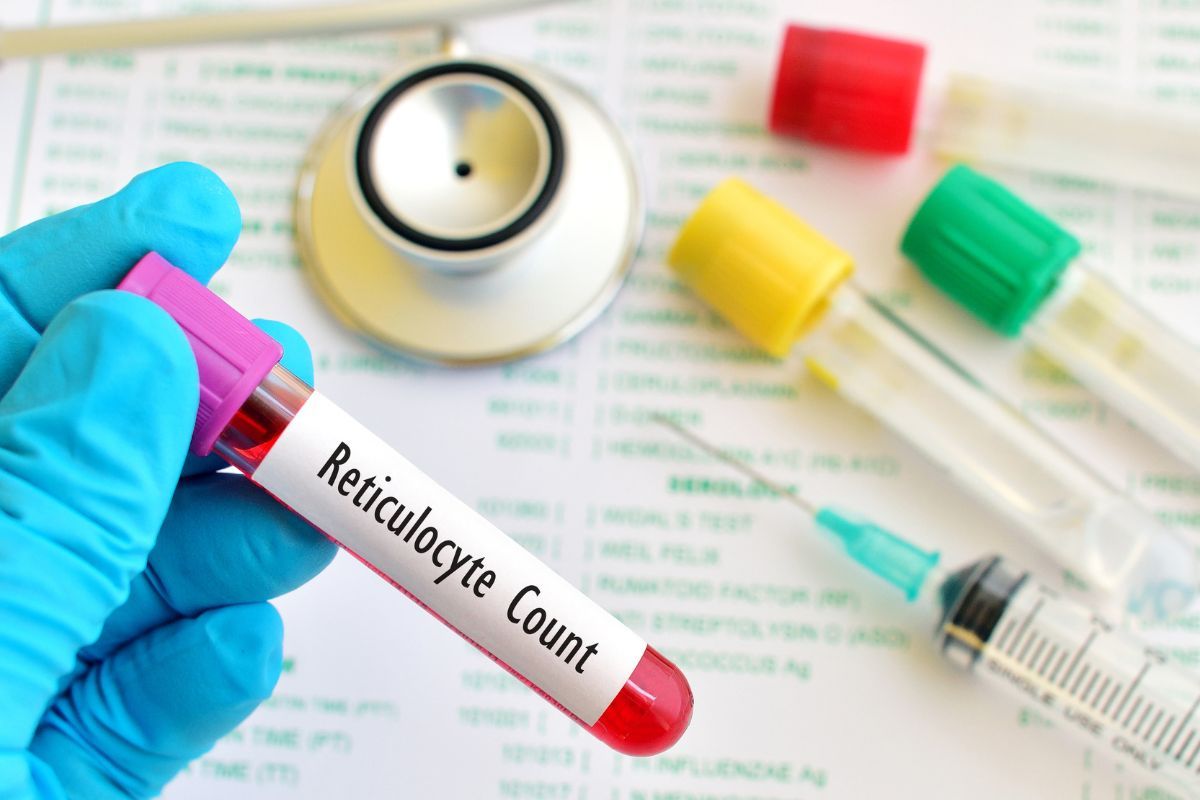
Blood morphology is one of the most commonly ordered diagnostic tests, which involves a detailed quantitative and qualitative analysis of the formed elements present in the blood. A sample of venous blood—usually taken from the antecubital vein—is collected and assessed using automatic laboratory analyzers. Morphology, also called a complete blood count, provides valuable information about the number, size, volume, and function of individual blood cells, such as erythrocytes (red blood cells), leukocytes (white blood cells), or thrombocytes (platelets). The test also allows the evaluation of indices such as hematocrit (HCT), hemoglobin concentration (HGB), or mean corpuscular volume (MCV). Blood morphology is a fundamental tool in diagnosing anemia, inflammatory conditions, bacterial and viral infections, coagulation disorders, or cancers. Thanks to this, it is one of the first tests doctors order, both in cases of suspected illness and during routine health screenings, and its results are often crucial to initiating appropriate treatment or further diagnostics.
Performing a blood morphology test is recommended in many different health situations and is part of the basic set of screening tests that everyone should undertake regularly, even if no symptoms are present. Routine blood morphology enables the early detection of abnormalities before clinical symptoms occur—especially in people exposed to stress, chronic fatigue, pregnant women, children, seniors, or those suffering from chronic diseases such as diabetes, hypertension, or autoimmune diseases. Doctors also often recommend the test in the case of symptoms such as recurrent infections, weakness, pale skin, excessive bleeding, unexplained bruising, high fevers, or disturbing results from other laboratory tests. Blood morphology is also a standard procedure before planned surgery, during pharmacological treatment (e.g., while taking medications that may affect bone marrow function), and to monitor the effectiveness of therapy for chronic conditions and cancer treatment. Ordering this test is a daily medical practice as it allows continuous monitoring of the general condition of the body, assessing the patient’s response to treatment, and quickly detecting concerning changes in the hematopoietic system. A significant advantage is the rapid turnaround time for results and the non-invasive nature of the procedure, making blood morphology suitable for virtually all ages. Regular testing helps build a so-called “reference base” for interpreting subsequent results and enables accurate tracking of any deviations from the norm, which is particularly important in the diagnosis of chronic and oncological diseases, early detection of vitamin or mineral deficiencies, and monitoring of general health.
Basic blood morphology parameters – what do the abbreviations in the results mean?
Blood morphology results include a series of abbreviations that refer to the key quantitative and qualitative characteristics of blood components. The most important are: RBC (Red Blood Cells, erythrocytes), WBC (White Blood Cells, leukocytes), PLT (Platelets), HGB (Hemoglobin), HCT (Hematocrit), MCV (Mean Corpuscular Volume), MCH (Mean Corpuscular Hemoglobin), MCHC (Mean Corpuscular Hemoglobin Concentration), and RDW (Red cell Distribution Width, index of erythrocyte anisocytosis). Reference values may vary depending on the laboratory, so always interpret them in light of the given norms. RBC indicates the number of red blood cells per milliliter of blood—a decrease may signify anemia, hyperhydration, or bleeding, while an increase can result from dehydration or diseases such as polycythemia vera. WBC shows the number of leukocytes, the white blood cells responsible for immunity. Their elevation often accompanies bacterial infections, inflammation, or cancer; low levels may result from viral infections, immunosuppression, or bone marrow damage. PLT, or platelet count, determines the blood’s ability to clot—deficiency (thrombocytopenia) poses a bleeding risk, while an excess (thrombocytosis) predisposes to thrombosis.
For qualitative parameters, HGB refers to the concentration of hemoglobin, the protein that transports oxygen, and is a basic marker for anemia. A decrease in this value is one of the first signs of anemia, while high levels can be observed in chronic obstructive pulmonary disease, dehydration, or high red blood cell counts. HCT (hematocrit) is the ratio of the volume of formed elements (mainly erythrocytes) to the total blood volume—a low value indicates anemia or hyperhydration, while a high value suggests dehydration or polycythemia. MCV assesses the average volume of a red blood cell—low MCV suggests microcytic anemia (most commonly due to iron deficiency), high MCV points to macrocytic anemia (e.g., due to vitamin B12, folic acid deficiency, or liver disease). MCH and MCHC indicate, respectively, the average amount of hemoglobin in a red blood cell and its average concentration within this cell. Significant deviations in these parameters are valuable clues for differentiating types of anemia; for example, low MCH/MCHC occurs in iron deficiency anemia, while high values are present in vitamin B12 deficiency. RDW shows variability in red blood cell size (anisocytosis)—a high RDW means the blood contains red cells of very different sizes, which is characteristic for some mixed or post-treatment anemias, or after recovery from iron or vitamin deficiencies. Also important in interpreting results is the automatic or manual leukocyte differential, which includes abbreviations such as NEU (neutrophils), LYM (lymphocytes), MONO (monocytes), EOS (eosinophils), BASO (basophils)—the percentage and absolute numbers of these cells can help differentiate infections, autoimmune, or allergic conditions. Understanding these abbreviations and their values is crucial not just for doctors but also for patients, who increasingly have direct access to their test results and want to actively manage their health and early detection of abnormalities.
Reference ranges for key blood indicators: WBC, RBC, PLT, MCV and more
The interpretation of blood morphology results relies on comparing the obtained values to established laboratory reference ranges, which may slightly differ depending on age, sex, health condition, and the analytical method used. Key parameters include WBC (white blood cells – leukocytes), RBC (red blood cells – erythrocytes), and PLT (platelets). WBC reference values in adults generally range from 4.0 to 10.0 x 10⁹/L. Values above this may indicate inflammation, bacterial infection, stress, or allergic reactions, while a decrease in WBC is seen in, among others, viral infections, bone marrow damage, or certain chronic autoimmune diseases. RBC values for women should be between 3.5–5.2 x 10¹²/L, and for men 4.2–5.7 x 10¹²/L. Too few red blood cells is typical for anemia and bleeding, while elevated RBC can occur with chronic hypoxia, dehydration, or chronic respiratory diseases. PLT reference ranges are generally 150–400 x 10⁹/L; decreased platelets (thrombocytopenia) increase the risk of bleeding, while exceeding the upper physiological range suggests thrombosis or ongoing inflammation, and can also be the response to some medications or infections. For HGB (hemoglobin concentration), the standard is 12–16 g/dL for women and 14–18 g/dL for men. Hemoglobin deficiencies are a classic marker of anemia, while high levels can be linked to chronic hypoxia or dehydration. Hematocrit (HCT) expresses the percentage of the volume of red blood cells relative to the total blood volume; reference ranges are 37–47% for women and 40–54% for men.
Additional diagnostic parameters provide a detailed characterization of red blood cells and hemoglobin. MCV (mean corpuscular volume) determines the average volume of a red blood cell, and its reference value is 80–97 fl. Decreased MCV is usually associated with microcytic anemia—most often due to iron deficiency or chronic inflammatory diseases. MCH (mean corpuscular hemoglobin) reflects the average mass of hemoglobin in a single erythrocyte, ranging from 27–33 pg—decreased MCH occurs in hypochromic anemia, and elevated MCH may indicate macrocytosis. MCHC (mean corpuscular hemoglobin concentration) should be 31–37 g/dL; low MCHC is typical for hypochromic anemia, while above-normal values are rare and may be caused by changes in cell membrane structure or measurement errors. RDW (red cell distribution width) indicates the range of size variation among erythrocytes—11.5–14.5% is normal, and deviations indicate heterogeneous populations of red blood cells, common in iron deficiency and mixed anemias. Morphology also includes a detailed white blood cell count (differential), giving percentage shares of lymphocytes (20–40%), neutrophils (50–70%), eosinophils (1–4%), basophils (0–1%), and monocytes (2–8%). Each deviation from these norms may have different clinical significance—for example, increased neutrophils typically indicate acute bacterial infection, while higher lymphocytes occur in viral or chronic inflammatory conditions. Reference ranges may also change in specific cases—during pregnancy, in children, elderly people, or athletes, so always interpret results in the individual’s context, symptoms, and medical history. Also, factors such as dehydration, stress, physical activity, recent surgery, or applied treatments can affect indicator values.
How to interpret blood morphology results in adults and children?
Interpreting blood morphology results requires consideration of many factors, including age, sex, current health status, and individual reference ranges set by laboratories. Standard reference ranges have been established for basic parameters in adults, but even small deviations do not always indicate disease—they are often temporary or the result of physiological states, diet, or medications. For instance, a mild drop in erythrocytes after intense exercise or in pregnancy is not always cause for concern. It is important to compare results to previous patient tests—sudden, significant changes may indicate acute pathology, while slow, gradual shifts often point to chronic processes. For adults, interpretation is mainly based on indices like RBC, WBC, PLT, HGB, HCT, as well as more advanced ones: MCV, MCH, MCHC, and RDW. An increase in leukocytes (WBC) above normal may indicate bacterial infection, inflammation, or a reaction to stress, while decreased values are frequently consequences of viral infection, autoimmune diseases, or nutritional deficiencies. Erythrocyte count and hemoglobin concentration are critical for diagnosing anemia—its types are differentiated using the size and hemoglobin content of red blood cells. Platelet count (PLT) is especially important—both deficiency (thrombocytopenia) and excess (thrombocytosis) may indicate serious hematologic conditions, inflammation, or medication side effects. In the elderly, chronically ill, or immunosuppressed, even minor changes in white blood cells can be clinically significant, so test results should always be interpreted in the broader context of the patient’s condition and clinical symptoms.
Special attention should be given to interpreting children’s blood morphology results, where reference ranges for parameters vary greatly depending on age and stage of development. Newborns, infants, preschool, and school-age children have their own dynamically changing norms, and correct ranges for parameters such as erythrocytes, leukocytes, or platelets may significantly differ from adult standards. For example, newborns naturally have higher hemoglobin and erythrocyte counts, which decrease over the first months of life, while leukocyte counts are usually higher than in adults and show wide variation. In infancy and childhood, drug resistance, viral infection, and iron deficiencies often present with atypical test fluctuations, so it’s always important to interpret results against specific norms for the child’s age and development stage. Also, children often temporarily exceed “adult” reference boundaries due to physiological events, like periodic white blood cell increases with infection or even strong emotional reactions during blood draws. Therefore, analysis of pediatric test results should include not only objective values but also the clinical picture and individual features of the child. Any abnormalities should be discussed with a pediatrician for proper interpretation. It’s crucial not to directly compare children’s values to adult standards, as this often leads to unnecessary concern. Evaluating children’s test results involves monitoring changes over time, assessing associated symptoms, and, if necessary, extending diagnostics to other lab or imaging tests. Any worrying or persistent deviations should always be clarified in cooperation with a physician, who will take into account the child’s entire clinical background and possible influencing factors.
Most common abnormalities and their possible causes
Abnormalities in blood morphology results are among the most frequently detected laboratory deviations and may be the first sign of developing health problems. Some of the most important parameters are the number of red blood cells (RBC), hemoglobin (HGB), and hematocrit (HCT). Decreased values indicate anemia, most commonly due to iron deficiency, chronic bleeding (e.g., from the gastrointestinal tract, heavy menstrual periods), chronic diseases such as kidney failure, and malabsorption of vitamin B12 or folic acid. Anemia can also accompany infections or hematological malignancies. Elevated RBC, HGB, and HCT can be seen in people living at high altitudes, smokers, those with chronic lung diseases, and in dehydration when the body compensates for lack of oxygen with increased red blood cell production or there’s a relative increase due to fluid loss. High indices can also result from polycythemia vera—a proliferative bone marrow disease. Changes in the number and proportion of white blood cells (WBC and different leukocyte subtypes) provide valuable information about the immune status. The most common abnormality is leukocytosis, or increased WBC above normal, indicating acute bacterial infection, inflammation, trauma, heavy exercise, stress, burns, or cancers such as leukemia. Dramatic rises in lymphocytes are found with viral infections (e.g., mononucleosis, flu), while neutrophilia is characteristic of bacterial infections or sepsis. Conversely, leukopenia, or a drop in white blood cells, is often caused by viral infection, nutrient deficiencies, treatment side effects (e.g., chemotherapy, immunosuppressive drugs), autoimmune diseases, or bone marrow damage. A seriously abnormal blood smear, showing the proportions of different leukocyte types (neutrophilia, lymphocytosis, eosinophilia, basophilia, monocytopenia), can be a key diagnostic clue for certain conditions; for example, eosinophilia is linked to allergies, parasites, or skin diseases.
Another frequently assessed parameter is platelet count (PLT), which is crucial for proper clotting function. Thrombocytopenia (low platelet count) is a serious bleeding threat and can be a result of viral infections (e.g., mononucleosis, flu virus), vitamin deficiencies (mainly B12 and folic acid), autoimmune diseases (idiopathic thrombocytopenic purpura), and sometimes drug complications or chemotherapy. It’s often seen in pregnant women in their third trimester, as well as in liver and spleen diseases or septic infections. On the other hand, thrombocytosis (high platelet count) is most often a sign of chronic infection, inflammation, chronic iron deficiency, or excessive marrow production in myeloproliferative cancers. Equally important are changes in qualitative parameters of red blood cells, such as MCV, MCH, MCHC, and RDW (variation in erythrocyte volume). Low MCV usually means microcytic anemia (iron deficiency), while high values indicate macrocytic anemia (B12, folic acid deficiency or liver diseases). A high RDW suggests the presence of cells of various sizes—typical in mixed anemias and during regeneration after treatment. Abnormal blood morphology can also occur in completely healthy people due to stress, dehydration, fatigue, or intense physical activity. That’s why results should always be interpreted considering the patient’s overall state, current symptoms, history, and medications, and any deviation from the norm should be consulted with a doctor, who may order further tests or initiate proper treatment.
When to consult a doctor about your blood test results?
Receiving blood morphology results often raises questions about interpretation and further action, especially when the results differ from established reference ranges. Consulting a physician is strongly recommended if the results show significant deviations from the norm—either above or below. Although minor differences may relate to temporary factors such as stress, physical exertion, dehydration, medications, or even the menstrual cycle, more serious or persistent changes require a broader analysis. Particular caution is needed with sudden, large fluctuations in basic parameters, such as a drastic drop in hemoglobin, platelets, or white blood cells, which may indicate a developing hematologic disease (such as anemia, leukemia, thrombocytopenia), immune disorders, severe infections, or chronic conditions. Alarming results also include a visible increase in blood parameters—for example, increased leukocytes may suggest an acute bacterial infection, inflammation, or, less commonly, hemopoietic system malignancies; excessively high erythrocyte counts or hematocrit may relate to chronic hypoxia, lung diseases, or abnormal blood cell production. Consulting a doctor is essential if serious conditions are suspected, especially if the abnormal results are accompanied by warning symptoms—pronounced fatigue, pale skin, breathing difficulties, loss of consciousness, frequent nosebleeds, unexplained bruising, fevers of unknown origin, or enlarged lymph nodes. In such cases, the doctor will not only assess if more in-depth diagnostics are needed (e.g., manual blood smear, biochemical or imaging studies) but also initiate appropriate treatment or refer to a specialist, such as a hematologist, oncologist, or immunologist.
It’s worth remembering that self-interpretation—even with online access to results—is difficult and risky for most patients. Decisions about consultation should be guided not only by abnormal parameters but also by general well-being, the emergence of new or worsening symptoms, and trends in results over time. Extra caution should be taken with children, pregnant women, the elderly, and those with chronic illnesses—abnormalities in these groups may quickly lead to serious complications. Not all deviations are a sign of disease; some are temporary or clinically insignificant. However, any situation causing doubts or concern should be explained with a healthcare provider, who will consider the patient’s individual characteristics, full clinical picture, and up-to-date medical guidelines. This ensures not only proper health assessment but also early detection of abnormalities, which may not produce typical symptoms at first. Consulting your results also provides information about the need for further tests, dietary or lifestyle changes, or specialist care—especially for risk groups: oncology patients, people with chronic kidney disease, cardiovascular disease, or women planning pregnancy. Working with your doctor ensures safety and allows you to consciously manage your health, minimizing risks and enabling rapid treatment, which is often crucial for effectiveness.
Summary
Blood morphology is one of the basic diagnostic tests, helping to detect many diseases and disorders early. Reading test results requires knowledge of reference ranges and interpreting key parameters such as WBC, RBC, PLT and MCV. Any deviations from the norm should be noted—they may indicate infection, inflammation, or hematological disorders. The final diagnosis is always made by a physician, but basic knowledge of blood morphology helps you take care of your own health and react quickly if concerning symptoms arise.